PhD: Multimodal AI for Ovarian Cancer Detection • Dr. Stéphanie Nougaret
3 years • 2025 • IRCM Montpellier
Defense planned for 2028
🩺 Medical Image Registration
🧫 MRI / WSI
🤖 Deep Learning
🖥️ VoxelMorph / Elastix
🔬 Virtual Biopsy
📊 Multimodal Fusion
Mission:
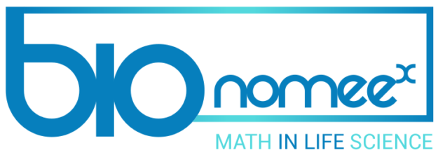
Building a virtual biopsy system for ovarian cancer — a modular framework that registers
high-resolution ex-vivo MRI (9.4T) with histopathological Whole Slide Images (WSI), enabling
biological feature maps (transcriptomics, carcinomatosis) to be projected onto
non-invasive MRI for clinical use.
Key Work:
- Modular Registration Pipeline: Sequential composition of global rigid/affine alignment (Elastix RRA) followed by B-spline local deformations and VoxelMorph deep learning dense flow fields — coarse-to-fine refinement of extreme non-linear tissue deformations.
- Custom Evaluation Framework: Two novel tissue-based metrics beyond standard intensity measures — area-based (Dice, Jaccard, Hausdorff) and landmark-based (expert pathologist-annotated anatomical keypoints), yielding clinically meaningful spatial validation.
- Annotation Software: Built a side-by-side annotation tool for expert pathologists to manually define biologically relevant MRI–WSI landmark.
Impact:
- First modular MRI–WSI registration framework validated for ovarian cancer.
- Enables transfer of biological feature maps from invasive WSI to non-invasive MRI coordinate space.
- Foundation for future clinical virtual biopsy tools — reducing reliance on invasive tissue sampling.
Next: Complete landmark annotation for the full IRCM cohort; extend to clinical 3T whole-body MRI; explore Virtual Biopsy deep learning models for direct biological feature prediction from MRI.